
| Overview | Cell Structures | Cell Migration | Cell Division |
Movies of Cell Division and Microtubule Dynamics during Cell Division |
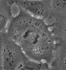 |
Phase Contrast Movie of a Dividing Normal Rat Kidney Cell |
This sequence starts with chromosomes already congregated into the "metaphase plate". Upon anaphase onset, the chromosomes are separated into two groups. The cell then divides into two daughter cells, each with one set of chromosomes. The movie ends with the decondensation of chromosomes and reformation of the nuclear envelope. Recording time, 19 min. |
|
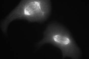 |
Microtubule Dynamics during Cell Division |
Microtubules are the main structural component of the mitotic spindle. They guide the movement of chromosomes and the transmission of signals for cytokinesis. Two cells here were microinjected during prometaphase with rhodamine-labeled tubulin, which became incorporated into spindle microtubules. After chromosomal separation, a set of midzone microtubules form in the central region and subsequently condense into bright bundles. Cytokinesis takes place along a plane defined by these midzone microtubule bundles. Toward the end of division midzone microtubule bundles condense into a bright midbody. Recording time, 28 min. |