
| Overview | Cell Structures | Cell Migration | Cell Division |
Dynamics of Actin Filaments during Early Cytokinesis |
Cao and Wang, J. Cell Biol. 111:1905-1911 (1990) |
To examine the movement of actin filaments during cell division, fragments of actin filaments labeled with fluorescent phalloidin, or fluorescent phalloidin itself, were microinjected into living Normal Rat Kidney cells. |
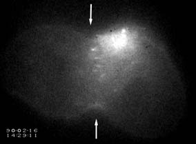 |
Movement of Microinjected Fragments of Actin Filaments into the Equatorial Region |
Actin filaments were assembled in vitro and labeled/stabilized with rhodamine phalloidin. The filaments were sheared into short fragments before microinjection into NRK cells during early anaphase. The site of injection appeared as a large bright spot near the top of the image. During the early phase of cytokinesis, several small aggregates of actin filaments dissociated from the site of injection and moved toward the equator, which is marked by two arrows. Recording time, 6 min. |
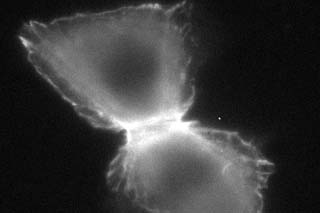 |
Movement and Contraction of Phalloidin-Labeled Endogenous Actin Filaments during Cytokinesis |
A well spread, dividing NRK cell was microinjected with fluorescent phalloidin to label its actin filaments. Filament bundles concentrate along the equator and radiate out. The movement of actin filaments during cleavage appears to involve both constriction along the equator and tearing in opposite directions toward the two poles. The structure disassembles at the same time. Recording time, 10 min. |